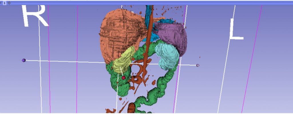
The majority of the organs were labelled and segmented from the VHP Data using 3dSlicer.
Figure 1.0: Segmenting the organs using 3DSlicer.
The segmented models were then sculpted and retopologized in Zbrush.
Figure 1.1: Sculpting the Lungs in ZBrush
Other organs, such as the intestines, that proved difficult to segment from the VHP Data, were modelled from scratch using splines in 3DStudio Max.

Figure 1.2: Modelling small intestines using splines in 3dsmax.
Next Step: The modelling of the organs was completed, and these organs were unwrapped and textured.

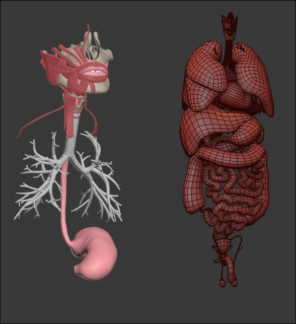
Figure 1.3: Organ modelling progress (left) and complete organ meshes (right).


